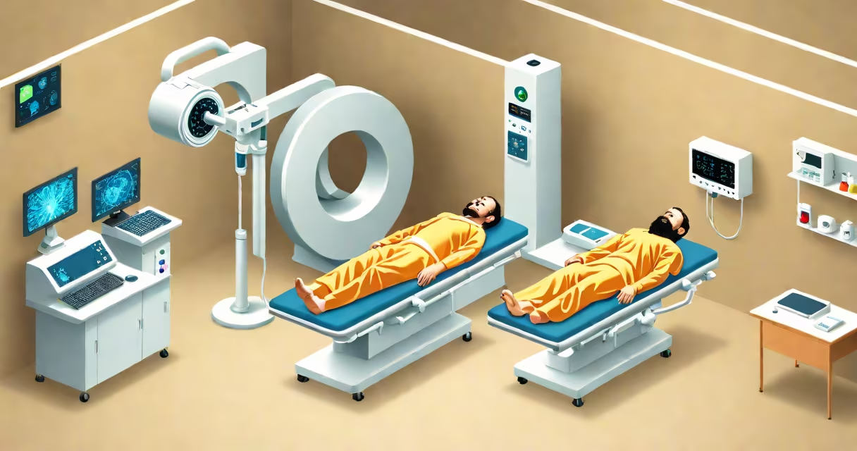
CT scans use X-rays and computer processing to create detailed cross-sectional images of internal structures. These scans help diagnose injuries, diseases, and tumors by providing clearer views than traditional X-rays.
A CT scanner rotates around the patient, taking multiple X-ray images from different angles. A computer combines these images to produce cross-sectional slices, or "tomograms," revealing bones, organs, and tissues in high detail. The process is painless and typically takes 10-30 minutes.
| Type | Focus Area | Common Uses |
|---|---|---|
| Head CT | Brain, skull | Stroke, trauma, tumors |
| Chest CT | Lungs, heart | Cancer, infections, blood clots |
| Abdominal CT | Organs, blood vessels | Organ damage, cancer, kidney stones |
| Spine CT | Vertebrae, spinal cord | Fractures, herniated disks |
Unlike MRIs, which use magnetic fields and radio waves, CT scans rely on X-rays. CT scans are faster and better for bone and emergency imaging, while MRIs provide superior soft-tissue contrast. A CT scan exposes patients to ionizing radiation, whereas MRIs do not.
Many people believe CT scans are always safer than X-rays. However, CT scans deliver higher radiation doses due to their detailed imaging. While the risk is low, repeated exposure increases cancer risk slightly. Doctors weigh benefits against risks, often using CT scans only when necessary.
During a car accident, a patient arrives at the ER with head trauma. A CT scan quickly reveals a brain hemorrhage, allowing doctors to act fast. Without the scan's speed and precision, diagnosing internal injuries would be much harder.
CT scans save lives by detecting critical conditions early. Their ability to show bones, organs, and blood vessels in 3D makes them indispensable in emergencies and complex diagnoses. Advances in technology continue to improve image quality while reducing radiation exposure.