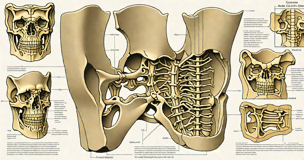
The median sacral crest is a bony ridge running along the midline of the sacrum, formed by fused vertebral bodies. It serves as a key landmark for spinal alignment and medical procedures.
The sacrum consists of five fused vertebrae, forming a triangular bone at the base of the spine. The median sacral crest runs along the posterior surface of this fusion, created by the overlapping spinous processes of these vertebrae. This ridge is easily palpable through the skin, making it a reliable external landmark.
The median sacral crest plays several critical roles:
Many people confuse the median sacral crest with the posterior sacral foramina, which are openings on either side of the crest. Unlike the foramina, the crest is a solid bony ridge, not a hollow space. Another common mistake is assuming the crest extends beyond the sacrum onto the coccyx. While the coccyx may have a similar ridge, it is not part of the median sacral crest.
In medical practice, the median sacral crest is important for several procedures. For instance, during a sacroiliac joint injection, physicians use the crest to locate the correct insertion point., in cases of spinal trauma, the presence or absence of the crest in imaging can indicate fractures or dislocations. According to the American Academy of Orthopaedic Surgeons, accurate identification of this landmark reduces complications during spinal surgeries.
I once observed a spinal surgery where the median sacral crest was used to align a patient's sacrum before inserting screws. The surgeon emphasized its importance in ensuring proper placement, highlighting how even a small misalignment could lead to serious complications.
The median sacral crest is a important anatomical feature that aids in spinal alignment, surgical precision, and diagnostic accuracy. Understanding its structure and function is essential for healthcare professionals and anyone studying human anatomy.