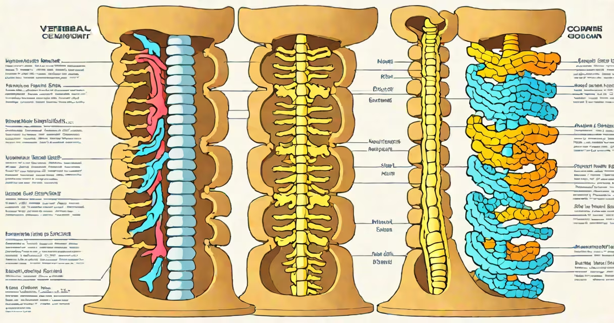
A vertebral column diagram illustrates the 33 stacked bones forming your spine, divided into five distinct regions: cervical (7 vertebrae), thoracic (12), lumbar (5), sacral (5 fused), and coccygeal (4 fused). This structure supports your body, protects the spinal cord, and enables flexible movement.
The vertebral column acts as a shock absorber while allowing rotation, flexion, and extension. Each vertebra connects via intervertebral discs (cartilage pads) that cushion impacts and maintain spacing. The spinal cord runs through the central canal formed by vertebral arches, branching into nerves that control bodily functions.
| Region | Vertebrae Count | Key Features |
|---|---|---|
| Cervical | 7 (C1-C7) | Smallest vertebrae; C1 (atlas) supports the skull; C2 (axis) enables head rotation |
| Thoracic | 12 (T1-T12) | Medium-sized; attach to ribs; limited mobility due to rib connections |
| Lumbar | 5 (L1-L5) | Largest vertebrae; bear most weight; prone to herniation |
| Sacral | 5 (fused as S1) | Forms the sacrum; connects to pelvis; part of the posterior pelvic wall |
| Coccygeal | 4 (fused as Co1) | Rudimentary tailbone; vestigial structure |
Many assume the spine has 31 bones, overlooking the fused sacral and coccygeal vertebrae. Others confuse vertebrae with ribs-though both protect organs, ribs attach to thoracic vertebrae while vertebrae form the spine itself. A vertebral column diagram clarifies these distinctions visually.
When diagnosing scoliosis, doctors compare patient X-rays to standard vertebral column diagrams. Deviations from the normal S-shaped curvature indicate lateral spine curvature. For instance, a 15° deviation from the thoracic kyphosis (normal inward curve) suggests mild scoliosis, requiring monitoring or treatment.