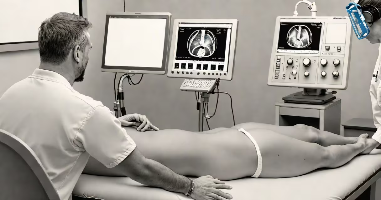
A bachelor of science in diagnostic medical sonography is a four-year undergraduate degree that trains students to perform ultrasound examinations, analyze images, and assist physicians in diagnosing medical conditions. This program combines coursework in anatomy, physiology, and patient care with hands-on clinical training.
The degree typically requires 120 credit hours, including general education courses, core sonography classes, and clinical rotations. Students learn to operate ultrasound equipment, interpret images, and communicate findings to healthcare providers. Clinical rotations provide real-world experience in hospitals, clinics, or imaging centers.
Most programs are accredited by the Commission on Accreditation of Allied Health Education Programs (CAAHEP), ensuring they meet industry standards. Graduates are eligible to take the American Registry for Diagnostic Medical Sonography (ARDMS) certification exams.
While the bachelor's degree covers general sonography, many programs offer specializations:
| Specialization | Focus Area | Common Procedures |
|---|---|---|
| Abdominal Sonography | Organs in the abdomen | Liver, gallbladder, pancreas scans |
| Obstetric/Gynecologic Sonography | Reproductive system | Prenatal ultrasounds, pelvic scans |
| Cardiac Sonography | Heart and blood vessels | Echocardiograms, stress tests |
| Vascular Sonography | Blood flow and vessels | Carotid artery scans, venous Doppler |
Diagnostic medical sonographers work in hospitals, diagnostic imaging centers, physicians' offices, and mobile imaging services. The demand for sonographers is growing due to the increasing use of ultrasound as a safe, non-invasive diagnostic tool. According to the U.S. Bureau of Labor Statistics, employment of diagnostic medical sonographers is projected to grow 14%, much faster than the average for all occupations.
Sonographers play a critical role in diagnosing conditions such as heart disease, cancer, and pregnancy complications. Their work helps reduce the need for more invasive procedures like biopsies or surgeries.
Many people confuse diagnostic medical sonography with other imaging technologies like X-rays or MRIs. Unlike these methods, ultrasound uses high-frequency sound waves rather than radiation. It is particularly valuable for monitoring pregnancies and assessing soft tissues, which are harder to visualize with other imaging techniques.
Another misconception is that sonographers only work in obstetrics. While prenatal ultrasounds are a significant part of their work, sonographers also perform scans on various body parts, including the heart, abdomen, and blood vessels.
During my clinical rotation at a local hospital, I assisted in performing an echocardiogram on a patient with suspected heart disease. Using the ultrasound machine, I captured images of the patient's heart chambers and valves. The cardiologist reviewed the images to assess heart function and diagnose potential issues. This hands-on experience reinforced the importance of precise imaging and clear communication between the sonographer and physician.
After graduation, I took the ARDMS certification exam and passed on my first attempt. The bachelor's program provided the technical skills and clinical experience needed to excel in this field.