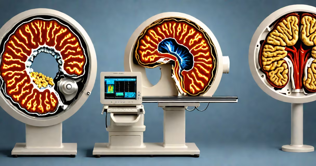
MRI (magnetic resonance imaging) and CT (computed tomography) scans are both medical imaging techniques, but they use different technologies to create images of the body. MRI uses useful magnets and radio waves to generate detailed images, while CT scans use X-rays to capture cross-sectional views.
MRI machines create a magnetic field around the body, aligning hydrogen atoms in the tissues. Radio waves disrupt this alignment, and the energy released as the atoms realign is measured to produce images. This process takes longer but provides superior contrast for soft tissues.
CT scanners rotate around the patient, sending X-ray beams through the body. Detectors measure the intensity of beams that pass through, creating cross-sectional images. This method is faster and better suited for bone and dense tissue imaging.
| Type | Common Uses |
|---|---|
| MRI | Brain and spinal cord imaging, joint and muscle evaluation, cancer diagnosis |
| CT | Head injuries, bone fractures, lung and chest issues, stroke diagnosis |
MRI is often preferred for examining soft tissues like the brain, spinal cord, and muscles. For example, it's the go-to for diagnosing torn ligaments or spinal disc issues. CT scans excel at capturing detailed images of bones, making them ideal for detecting fractures or assessing lung conditions.
Many people believe MRI is always safer because it doesn't use radiation. While true, MRI has its own risks, such as complications for patients with metal implants. CT scans, though involving radiation, are generally safe for most patients when used appropriately.
Another myth is that MRI provides better images. In reality, CT scans often offer faster results and clearer bone images, while MRI excels with soft tissues.
After a car accident, a patient might undergo a CT scan first to quickly check for bone fractures or internal bleeding. If the initial results suggest soft tissue damage, an MRI could follow to assess muscle or nerve injuries more precisely.