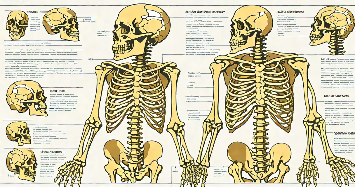
Skeletal anatomy and physiology is the study of bones, their structure, and how they function within the body. This system provides support, facilitates movement, protects internal organs, and produces blood cells.
The skeletal system consists of 206 bones in adults, connected by joints and supported by muscles and ligaments. Bones serve as levers for movement when muscles contract. They also store minerals like calcium and phosphorus and house bone marrow, which produces red and white blood cells.
Bones vary in shape and function:
| Type | Description | Examples |
|---|---|---|
| Long bones | Longer than wide, designed for strength and movement | Femur, humerus |
| Short bones | Cube-shaped, provide stability and support | Wrist and ankle bones |
| Flat bones | Thin and flat, protect organs and provide surface area for muscle attachment | Skull, ribs, sternum |
| Irregular bones | Various shapes, fit complex functions | Vertebrae, hip bones |
The skeletal system supports the body's weight and shape. It protects important organs like the brain (skull), heart (ribs), and spinal cord (vertebrae). Bones also act as a mineral reservoir, releasing calcium and phosphorus into the bloodstream when needed.
Bone marrow, found within bones, produces red blood cells, white blood cells, and platelets. This process, called hematopoiesis, is important for maintaining healthy blood and immune function.
Many people think bones are inert structures, but they are actually living tissues that constantly remodel themselves. Osteoclasts break down old bone, while osteoblasts build new bone. This process ensures bones remain strong and adapt to stress.
Another misconception is that bone density peaks in early adulthood and then declines. While it's true that bone density decreases with age, regular exercise and a diet rich in calcium and vitamin D can help maintain bone health.
The femur, or thighbone, is the longest and strongest bone in the body. It supports the body's weight and facilitates movement through its connection to the hip and knee joints. The femur's structure includes a shaft (diaphysis) and two ends (epiphyses) covered with articular cartilage for smooth movement.
During growth, the epiphyseal plate, a layer of cartilage, allows the bone to lengthen. Once growth is complete, this plate ossifies, turning into solid bone. Understanding the femur's anatomy and physiology helps in treating fractures and conditions like osteoarthritis.
While skeletal anatomy focuses on the structure and organization of bones, skeletal physiology examines how bones function. For example, anatomy studies the shape and connections of bones, while physiology investigates how bones grow, repair themselves, and interact with other systems like the muscular and endocrine systems.
Deer have unique skeletal adaptations for their environment. Their legs are long and slender, designed for running and jumping. The deer skeleton also includes antlers, which are not true bones but extensions of the skull covered in velvet during growth. Understanding deer skeletal anatomy provides insights into their behavior and survival strategies.