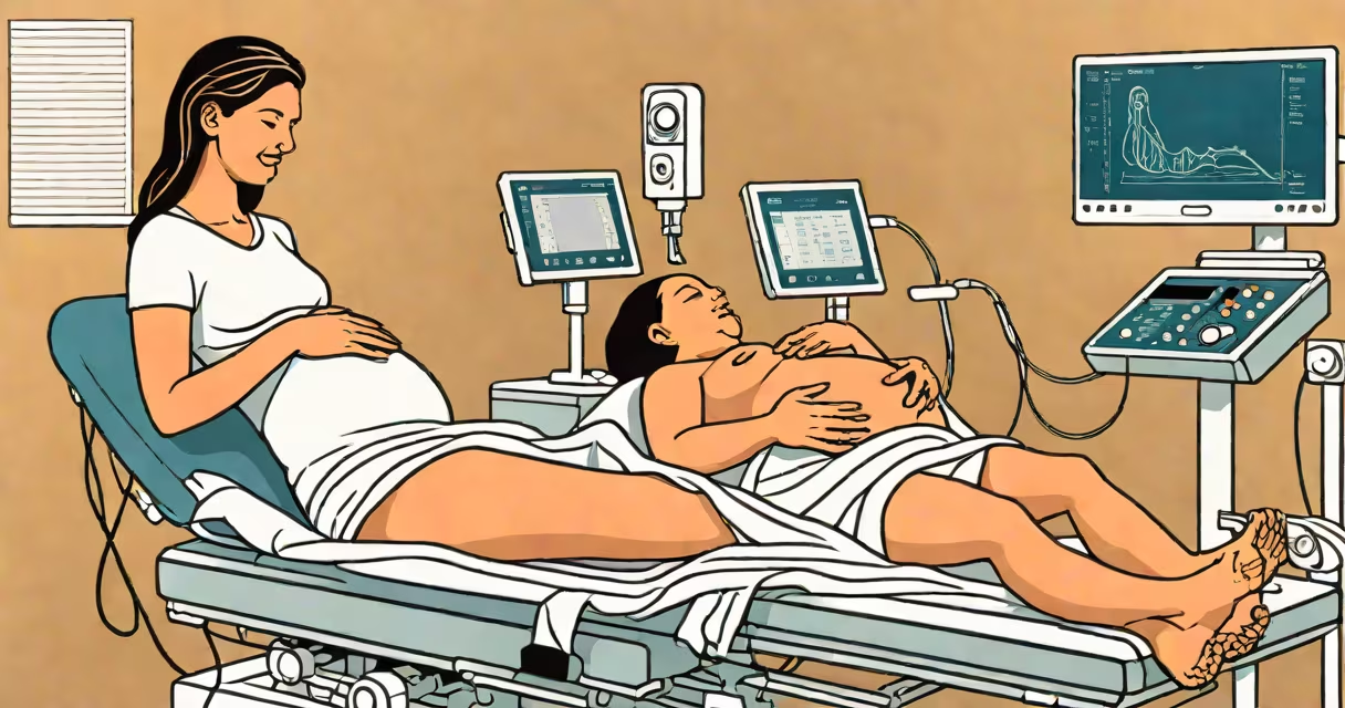
Your first ultrasound is a transvaginal or abdominal scan between 6-9 weeks to confirm pregnancy viability, check fetal heartbeat, and estimate due date.
Ultrasounds use high-frequency sound waves to create images of your uterus and developing embryo. A wand-like transducer emits sound waves that bounce back to create a visual image on a screen. The sonographer applies gel to your skin to help transmit the sound waves.
| Type | When It Occurs | Purpose |
|---|---|---|
| Transvaginal ultrasound | 6-9 weeks | Confirms viability, detects heartbeat, measures gestational sac |
| Abdominal ultrasound | 11-14 weeks | Nuchal translucency screening, estimates due date |
For a transvaginal ultrasound, you'll lie on your back with legs in stirrups. The sonographer inserts a lubricated probe into your vagina. For an abdominal ultrasound, you'll have a full bladder and lie on your back while the sonographer moves a transducer over your belly. Both procedures take about 15-30 minutes.
The sonographer will show you the gestational sac, yolk sac, and fetal pole on the screen. Around 6 weeks, you may hear the fetal heartbeat, which sounds like a rapid tapping. The sonographer will measure the embryo's size to estimate the due date.
Many people think the first ultrasound determines the baby's sex, but this usually isn't possible until later scans., some believe ultrasounds are painful, but they're generally comfortable aside from potential discomfort from a full bladder during abdominal ultrasounds.
If you're nervous about your first ultrasound, remember that it's a routine part of prenatal care designed to ensure your pregnancy is progressing normally. The American College of Obstetricians and Gynecologists recommends at least one ultrasound during pregnancy, with additional scans depending on your healthcare provider's recommendations.