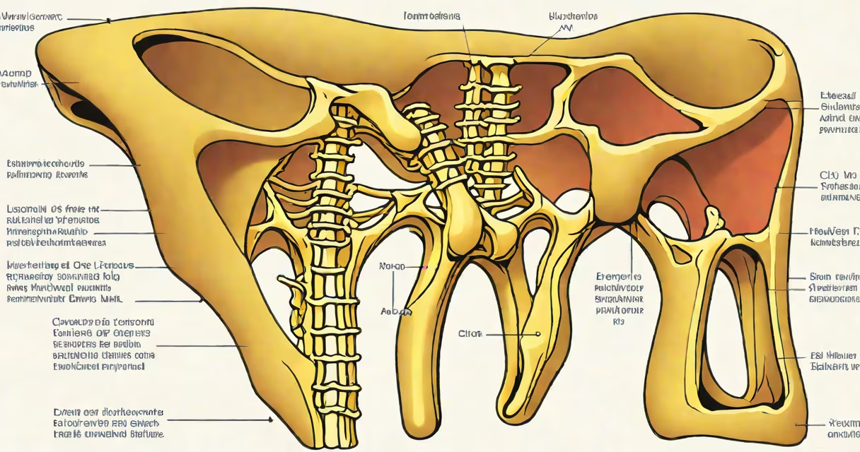
A hip bone diagram labeled shows the three fused bones forming the os coxae: the ilium, ischium, and pubis. These structures create part of the pelvis, supporting the spine and facilitating movement.
The hip bone connects to the sacrum at the back and to other hip bones at the front via the pubic symphysis. It forms the socket (acetabulum) for the femoral head, creating the ball-and-socket hip joint. This structure allows for many motion while bearing significant weight.
The hip bone consists of three primary components:
The acetabulum, formed by all three bones, is the key landmark for hip joint function.
Doctors use labeled diagrams to explain injuries, such as fractures or arthritis, to patients. Physical therapists reference them when designing rehabilitation programs. Surgeons rely on detailed diagrams during procedures like hip replacements.
Many people confuse the hip bone with the entire pelvis or assume it's a single bone. In reality, each hip bone is a fusion of three separate bones that develop during childhood and fuse by adulthood. Another mistake is thinking the hip joint is primarily for weight-bearing; it also enables complex movements like rotation and flexion.
For instance, the ischium is often overlooked in favor of the ilium, yet it bears most of the body's weight when sitting. Understanding this distinction is important for treating conditions like sciatica, which involves pressure on the ischial tuberosity.
Consider a patient with a fractured pubic ramus. A labeled diagram highlights the specific area of injury, helping clinicians assess damage to nearby structures like the bladder or groin muscles. The diagram also guides treatment, such as whether surgery or physical therapy is needed.
For further study, consult anatomical resources like Gray's Anatomy or medical textbooks specializing in orthopedics. These references provide detailed illustrations and explanations of hip bone anatomy and related conditions.