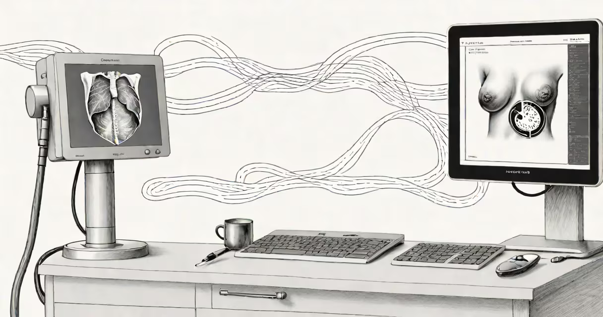
A breast sonogram is an ultrasound imaging test that creates pictures of breast tissue using high-frequency sound waves. Doctors often use it to evaluate changes seen on a mammogram or to investigate lumps felt during a clinical breast exam.
During the procedure, a technician applies a special gel to the breast and then moves a handheld device called a transducer over the skin. The transducer emits sound waves that echo back when they hit dense tissue. A computer converts these echoes into black-and-white images displayed on a monitor.
The entire process takes about 15-30 minutes and is painless. Unlike mammograms, which use ionizing radiation, sonograms use only sound waves, making them safe for repeated use.
| Type | Primary Use | When Ordered |
|---|---|---|
| Diagnostic ultrasound | Evaluates breast lumps or abnormalities | After an abnormal mammogram or physical exam |
| Breast ultrasound biopsy | Guides needle biopsies of suspicious areas | When a lump needs tissue sampling |
| Automated breast ultrasound (ABUS) | Screening tool for dense breast tissue | For women with dense breasts and normal mammograms |
Many people confuse breast sonograms with mammograms. While both create images of breast tissue, they use different technologies. Mammograms use low-dose X-rays to detect tumors, while sonograms use sound waves to examine tissue structure.
Another misconception is that sonograms replace mammograms. They don't. The American Cancer Society recommends mammograms as the primary screening tool, with sonograms used for further evaluation when needed.
Doctors typically order a breast sonogram when a mammogram shows an unclear area or when a lump is felt but doesn't appear on a mammogram. Sonograms are particularly useful for women with dense breast tissue, as mammograms may miss some abnormalities in denser tissue.
Sonograms can also help determine whether a lump is solid or filled with fluid (a cyst). If the lump is solid, further testing, such as a biopsy, may be needed. If it's a cyst, no additional testing is usually required unless symptoms change.
In my experience working with imaging centers, I've seen sonograms provide clarity in cases where mammograms were inconclusive. For example, a patient with dense breast tissue and a palpable lump had a normal mammogram, but the sonogram revealed a small cyst that resolved on its own without intervention.
While sonograms are excellent for evaluating specific areas, they aren't as effective as mammograms for screening the entire breast. They also have limitations in detecting small calcifications, which are often early signs of cancer. That's why sonograms are used alongside mammograms, not as a replacement.
According to the Mayo Clinic, sonograms are also operator-dependent, meaning the quality of the images relies heavily on the technician's skill and experience. Proper training ensures accurate results.
A breast sonogram is a valuable diagnostic tool that complements mammograms by providing detailed images of breast tissue using sound waves. It helps clarify abnormalities, guide biopsies, and monitor changes in breast tissue. While it doesn't replace mammograms, it is important for breast cancer screening and diagnosis.
For more details on breast health, including screening guidelines, visit the breast health guide.