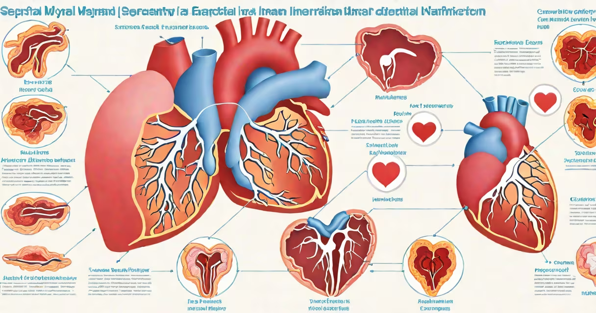
Septal myocardial infarction is a heart attack damaging the interventricular septum, the wall separating the heart's left and right ventricles. This type often occurs alongside anterior wall infarctions due to shared blood supply.
The heart's septum prevents oxygen-rich and oxygen-poor blood from mixing. When a septal infarction occurs, blood flow to this area is blocked, typically by a coronary artery clot. The damage can disrupt heart function, leading to arrhythmias or heart failure if untreated.
Septal infarctions are categorized by location and severity:
| Type | Description | Common Causes |
|---|---|---|
| Anterior Septal Infarction | Affects the front portion of the septum | Blockage in the left anterior descending artery |
| Inferior Septal Infarction | Affects the lower part of the septum | Blockage in the posterior descending artery |
Anterior septal infarctions are more common and severe due to the larger muscle mass involved. Inferior septal infarctions may cause fewer symptoms initially but still require urgent care.
Septal infarctions are diagnosed using ECG patterns, echocardiograms, and cardiac enzymes. Treatment includes clot-busting drugs, angioplasty, or surgery to restore blood flow. Early intervention reduces long-term damage.
One non-obvious fact: septal infarctions can lead to ventricular septal rupture, a life-threatening complication where the septum tears. This occurs in about 0.2% of cases but requires immediate surgical repair.
Many confuse septal infarctions with general heart attacks. While all heart attacks involve muscle damage, septal infarctions specifically target the septum. Another mistake is assuming only anterior wall heart attacks are dangerous-inferior wall infarctions can also be fatal.
For example, a patient with chest pain and an ECG showing ST elevation in leads V1-V4 likely has an anterior septal infarction. Treatment would focus on restoring blood flow to the left anterior descending artery.
During my time working in a cardiac unit, I observed a patient with an inferior septal infarction. Their ECG showed changes in leads II, III, and aVF, indicating blockage in the posterior descending artery. Prompt treatment with angioplasty saved their life, highlighting the importance of rapid diagnosis.