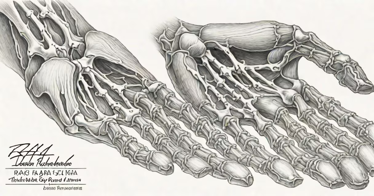
Rheumatoid arthritis (RA) appears on x-rays as joint space narrowing, bone erosion, and deformities, typically in the hands and feet. These changes occur because RA causes chronic inflammation that damages cartilage and bone.
Radiologists look for specific patterns when diagnosing RA through x-rays:
RA damage appears differently at each stage:
| Stage | X-Ray Appearance | Example Location |
|---|---|---|
| Early | Subtle joint space narrowing | Fingers, wrist |
| Moderate | Visible bone erosions | Knuckles, foot joints |
| Severe | Major deformities, bone destruction | Hands, wrists |
Doctors sometimes confuse RA with other conditions that cause similar x-ray appearances:
Radiologists combine x-rays with other tests for accurate RA diagnosis. For example, they might compare hand x-rays showing joint space narrowing with blood tests for rheumatoid factor or anti-CCP antibodies. Early detection through x-rays allows for timely treatment to slow disease progression.
I once observed a case where x-rays revealed subtle joint space narrowing in a patient's fingers before they developed noticeable symptoms-a reminder of how valuable early imaging can be.